Currently under study
The unique optical properties of gold nanoparticles make them attractive for a wide range of applications which require optical detection and manipulation techniques. Here, we experimentally demonstrate the use of single femtosecond pulses at resonance wavelength for a controlled conjugation of gold nanoparticles and fluorescent proteins. This optically driven reaction is rigorously studied and analyzed using a variety of experimental techniques, and a detailed model is proposed which describes the adsorption of the proteins onto the nanoparticles’ surface, as well as their subsequent desorption by a reducing agent. Potential applications of the resulting nanoparticle-protein conjugates include controlled delivery of fluorescent markers and local sensing of biochemical processes.
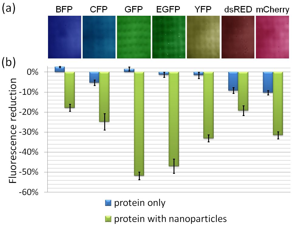
Figure 1
Fig. 1. (a) Fluorescence images of the nanoparticle-protein solutions following resonant illumination by a single pulse. The (false) colors correspond to the fluorescence emission wavelength of each protein. The field of view of each frame is 2.5 mm × 1.87 mm. (b) A bar chart showing the relative changes of the total fluorescence intensities after laser irradiation for protein-only solutions (blue bars) and for nanoparticle-protein solutions (green bars).

Figure 2
Fig. 2. Extinction spectra of illuminated nanoparticles solution with GFP before (dashed blue curve) and after a single pulse illumination (solid blue curve). Loss of the absorption peak after single pulse irradiation was observed when the nanoparticles were mixed with Myoglobin (solid black curve), Streptavidin (solid red curve), and without proteins (solid green curve). Left inset: TEM image of an irradiated nanoparticles-GFP solution. Right inset: TEM image of irradiated gold nanoparticles in the absence of fluorescent proteins. Scale bars represent 200 nm.
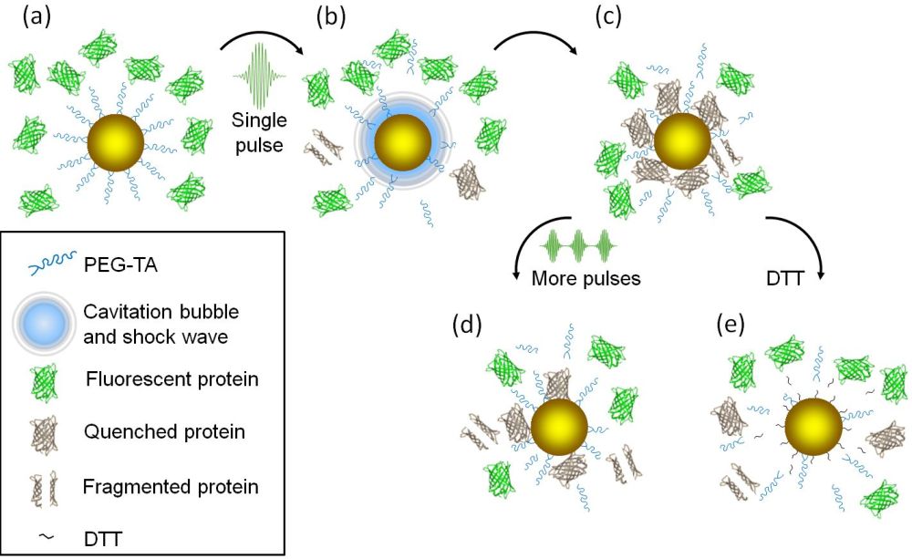
Figure 3
Fig. 3. Proposed model for optical pulse-driven adsorption of fluorescent proteins on gold nanoparticles. (a) Gold nanoparticles coated with PEG-TA within a solution of fluorescent proteins. (b) Irradiation by a single femtosecond pulse results in a cavitation bubble and shock wave formation, leading to partial removal of the PEG molecules. (c) The fluorescent proteins are adsorbed onto the particle and their fluorescence is quenched. (d) Following additional pulses, the quenched proteins are gradually degraded. (e) The addition of DTT to the solution results in protein desorption from the nanoparticles.
References
- Controlled fabrication of gold nanoparticles and fluorescent proteins conjugates
Gili Bisker, Limor Minai and Dvir Yelin
Plasmonics , (2012)