Gold nanoparticles find a wide range of applications in optics and photonics; however, their detailed interaction with intense laser light is only partially understood. Previous works have studied the effect of intense pulse trains on gold nanoparticles at a wide range of illumination parameters, and observed diverse optical and morphological changes. In this work we study, for the first time, the interaction between single femtosecond pulses and gold nanoparticles. Using transmission electron microcopy and optical spectroscopy, we have found that nanoparticles illuminated by 50 fs pulses with fluence of less than 0.15 J/cm2 per pulse (3 TW/cm2) undergo morphological changes which affect their extinction spectra. Experimentation with particles of different diameters show similar qualitative effects, which are more pronounced for larger particles. Pulses at different excitation wavelengths were found to induce different effects for resonance and off-resonance conditions. The presented results provide valuable experimental data on the complex pulse-particle interaction and would be helpful for better understanding of the physical processes that are involved.
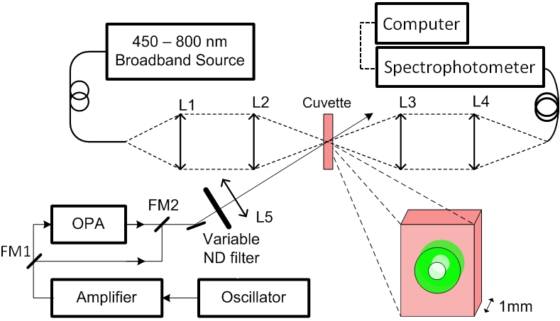
Figure 1
To study the effect of femtosecond pulses on the gold nanoparticles, nanoparticle solutione were placed in a 1 mm thick rectangular glass cuvette, which was placed at the focal plane of an optical arrangement (Figure 1). This experimental setup allowed the illumination of the particles with single pulses while observing changes in the extinction spectra. Light from a visible broadband source was coupled into a multimode optical fiber whose distal end was imaged into a small spot on the cuvette using two achromatic lenses L1 (75 mm focal length) and L2 (50 mm focal length). The focal depth was approximately 5 mm, longer than the total cuvette thickness (1 mm). The light transmitted through the cuvette was then collimated (lens L3, 75 mm focal length) and refocused (lens L4, 150 mm focal length) into a multimode fiber, which was used as the input of a commercial spectrophotometer having a maximal capturing rate of 250 spectra per second. The wavelength-dependent extinction coefficients were computed by taking the logarithm of the ratio between the transmitted spectra of pure water and that of the nanoparticle solution.
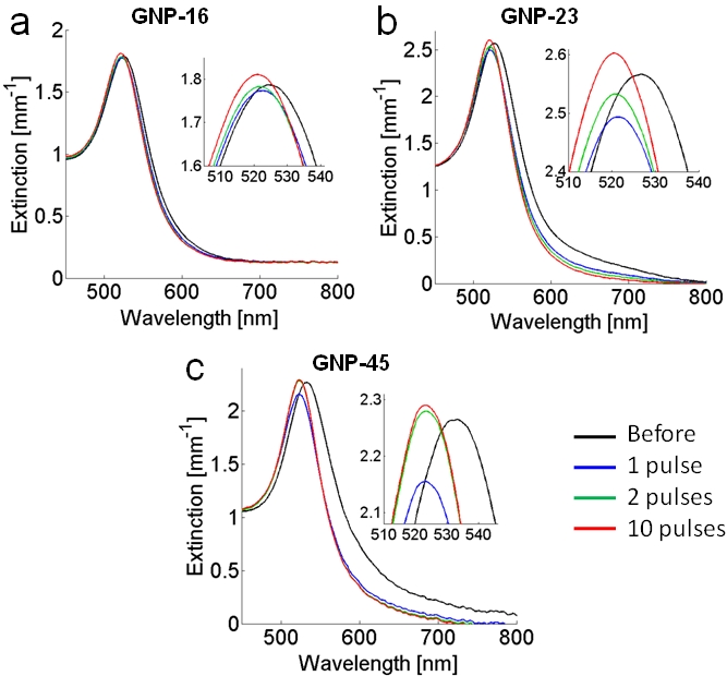
Figure 2
The extinction spectra of solutions having particle diameters of 16, 23, 45 nm following illumination of single 50 fs pulses of 90 mJ/cm^2 per pulse (1.8 TW/cm^2), at a center wavelength of 550 nm, are shown in parts a, b, and c respectively of Figure 2. A comparison between the extinction spectra before illumination (black line) and immediately after (less than a second) 1 pulse (blue), 2 pulses (green), and 10 pulses (red) reveal noticeable differences in the extinction spectra after each pulse. The effect of the first pulse was the most significant for all three solutions, and included noticeable blue shifts of the spectra, spectral narrowing by 5-25%, decrease in the maximum extinction values by approximately 5%, and a substantial decrease of the extinction at near-infrared wavelengths by 70-90%. The effect of the second pulse was less pronounced but had a similar trend, with the exception of the peak extinction values which slightly increased compared to the peak values after 1 pulse. Similar effects, although gradually decreasing in magnitudes, were observed after each additional pulse, until no changes were visible after more than 10 pulses.
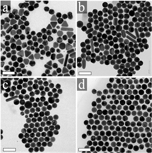
Figure 3
The effect of the resonant illumination at 550 nm could be noticed by comparing the TEM images of the GNP-45 solution before illumination (Figure 3a), after 1 pulse (3b), 2 pulses (3c), and 10 pulses (3d). Before illumination (3a), the solution contained mainly nanospheres, but also a certain quantity (approximately 20%) of different particle shapes, including triangles, rods, and icosahedrons. After the first pulse illumination, a more homogeneous assortment of particles was observed, with a smaller abundance of nanorods and of other particles with sharper corners or edges (3b). This trend continued with each additional pulse (3c,d), until almost all of the particles were transformed into spheres with a narrower size distribution. A similar effect was evident by visual examination of the TEM images of the resonantly illuminated drops from the GNP-23 and GNP-16 solutions (data not shown). Scale bars represent 100 nm.
References
- The effect of single femtosecond pulses on gold nanoparticles
Omri Warshavski, Limor Minai, Gili Bisker, Dvir Yelin
J. Phys. Chem. C 115, 3910 (2011)