Biomedical Optics Laboratory
Welcome to the Biomedical Optics Laboratory at the Technion. Our research involves several projects in clinical diagnosis and therapy, focusing on developing entirely novel and unique technologies. Our optical diagnosis work uses label-free in vivo microscopy for imaging various clinical conditions. For example, we have developed methods for imaging blood cells in rapid flow for painless, noninvasive blood count, as well as methods for mapping the nanometric vibrations of the tympanic membrane, aiming at effective functional diagnosis of various hearing problems. Our optical therapy work involves using gold nanoparticles and ultrashort laser pulses for triggering various effects in specifically targeted cells, including specific cell death for cancer therapy, and specific cell-cell fusion for tissue regeneration.
Needless to say, our group is constantly seeking for excellent, open minded students.
Diagnostics
Among all clinical imaging modalities, optical imaging provides the highest resolution and contrast. A main research effort in our group is the study and development of optical imaging techniques that are useful for clinical applications, including minimally-invasive endoscopic techniques with minimal mechanical scanning, low coherence interferometric imaging, and reflectance confocal microscopy.

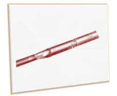
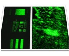
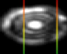
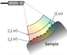
Utilizing Fourier-domain interferometry, spectrally encoded endoscopy (SEE) was shown capable of video-rate three-dimensional imaging…
Read more about adjusting field of view using dispersion
Therapy
Lasers are becoming increasingly popular for numerous medical applications. The use of femtosecond laser pulses is particularly attractive for such applications thanks to the “cold” process of tissue ablation which is confined only to the focal volume of the laser beam. In our group we use femtosecond pulses of various wavelength to excite specifically targeted noble-metal nanoparticles at their plasmonic resonance. The combined effect of the short pulses and small particles allows for specific manipulations of cells and tissue with minimal toxicity and collateral damage.
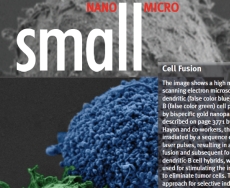
The attachment between two different cells via a bispecific nanoparticle is illustrated in Figure 1a. Following the addition of the nanoparticles…
Read more about specific cell fusion

Specifically targeting and manipulating living cells is a key challenge in biomedicine and in cancer research in particular…
Read more about nano-manipulations of cells
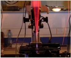
Gold nanoparticles find a wide range of applications in optics and photonics; however, their detailed interaction with intense laser light is only partially understood…
Read more about nanoparticle-pulse interaction
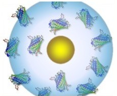
The unique optical properties of gold nanoparticles make them attractive for a wide range of applications which require optical detection and manipulation techniques…
Read more about nano-manipulations of macro-molecules

