In its current mode of implementation, SEE has several limiting factors which need to be addressed before its clinical promise could be realized. First, the use of wavelength to encode space imposes some difficulties on wavelength-sensitive imaging modalities. For example, fluorescence spectrally encoded imaging required a sophisticated optical setup for frequency-encoding [19]. Additionally, the use of spatially coherent illumination through a single mode fiber causes pronounced speckle noise, small depth of field, and poor signal collection efficiency which often requires the use of lasers, supercontinuum generation sources, or high power super-luminescent diode arrays. One possible solution for addressing these issues includes the use of a double-clad fiber for spatially coherent sample illumination and incoherent signal collection. While double-clad SEE was demonstrated capable of speckle-free imaging with large depth of field, the endoscopic probe itself suffered from significant cross-talk between the illumination and the collection channels. Back reflections from the probe’s optics, which were efficiently collected by the large area and the high numerical aperture of the inner cladding, resulted with high image noise and required continuous background subtraction during image acquisition.

Figure 1
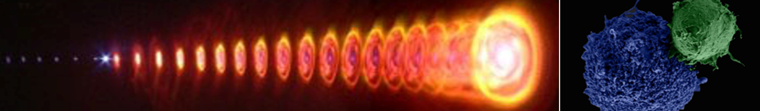
We show a new approach for SEE, termed multiple-channel SEE (MC-SEE), which addresses many of the image quality concerns of SEE and further expands its functionality. While current forms of SEE involve spectral encoding in both the collection and illumination channels independently, effective encoded imaging is still feasible using only one encoded channel. The concept of imaging with a single encoded channel is schematically illustrated in Fig. 1, showing space-to-wavelength encoding of broadband light (e.g. fluorescence) emanating from a specimen.

Figure 2
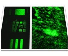
MC-SEE images of a portion of a 1 Euro cent coin using spectrally encoded coherent (top panel) and incoherent front illumination (bottom panel) are shown in Fig. 2. Comparison between the two images reveals significant reduction in speckle noise using incoherent illumination, resulting in a more natural appearance with better discrimination of surface texture.
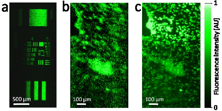
Figure 3
An image of a USAF-1951 fluorescence resolution target was acquired using incoherent excitation light (Nikon Fiber Illuminator, 130 W mercury lamp) and a 360-440 nm bandpass excitation filter, with exposure time of 100 ms per line (Fig. 3a). Along its wavelength (horizontal) axis, the fluorescence image revealed an intensity profile which resembles the emission spectrum of the fluorophore, resulting with a field of view of approximately 1.5 mm in the x axis. To demonstrate the potential of our approach to image weak fluorescence signals of biological specimen, we have imaged a culture of fixed human epithelial cells of breast adenocarcinoma origin (MDA-MB-231), whose membranes were labeled with a green fluorescent marker (DiOC18), using a 465-495 nm bandpass excitation filter. The resulting MC-SEE fluorescence image of the cells, acquired with 500 ms line exposure time, is shown in Fig. 3b, next to an image of the same field of view using a conventional epi-fluorescence microscope (NA = 0.45, Fig. 3c). While the numerical aperture of the SEE lens (NA = 0.25) and the signal-to-noise ratio (6.3 dB) were too low to resolve sub cellular structures, single cells could be easily resolved and a good match was obtained between the two images.
References
- Multiple-channel spectrally encoded imaging
Avraham Abramov, Limor Minai, and Dvir Yelin
Opt. Express 18, 14745(2010)